


Six-month journey to find treatment
Mr. Bohdan Vasiluk, 37 years old, a Ukrainian national living in Da Nang, went to the dentist after experiencing a toothache. An X-ray revealed a large cystic lesion in his lower jawbone.
Because the lesion was beyond the dentist’s scope of treatment, he was advised to seek care from a more specialized facility. For nearly six months, he visited numerous clinics and hospitals in Vietnam and even traveled to Thailand in search of treatment, but still could not find a suitable solution.
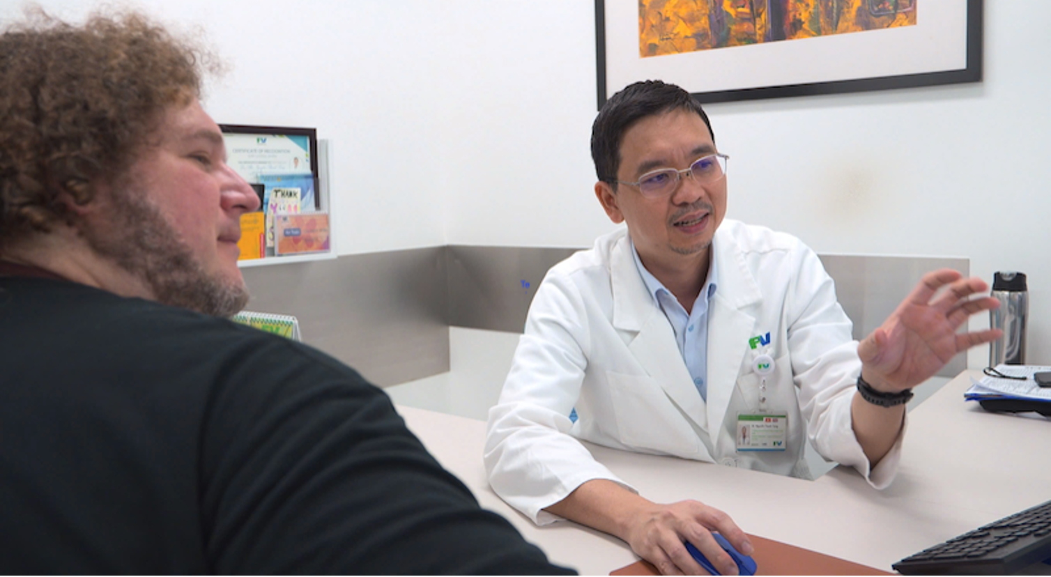
Mr. Vasiluk was later referred to FV Hospital by a Vietnamese doctor. He emailed the hospital with his medical history and previous images, and his medical records were then forwarded to Dr. Nguyen Thanh Tung, PhD, Head of the Maxillofacial and Dental Surgery Department at FV Hospital.
After reviewing the images, Dr. Tung told Mr. Vasiluk that his condition was treatable. After months of searching for an answer, the 37-year-old finally had a clear treatment plan.
Large cyst, but almost no symptoms
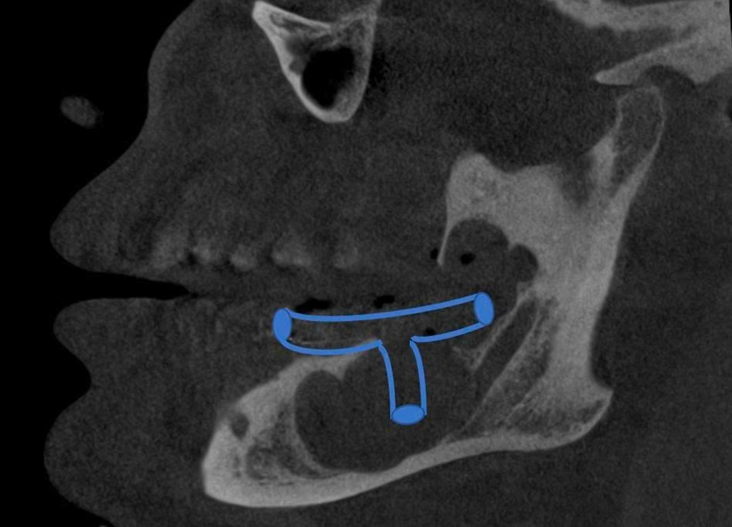
From the outside, Mr. Vasiluk’s face and oral cavity appeared almost normal. However, 3D reconstruction images showed a lesion approximately 56mm in diameter that had destroyed a large portion of the mandible and extended into the ascending ramus. The lesion had also displaced the mandibular canal and caused bone resorption around the roots of two left mandibular molars.
According to Dr. Tung, cystic lesions in the jawbone can progress silently. Some cases do not cause obvious pain or swelling and are only discovered incidentally when patients undergo dental X-rays.
Medical literature also indicates that jawbone cysts can grow to a considerable size before causing noticeable symptoms. When a lesion becomes extensive, treatment may need to take into account the risk of damage to the teeth, nerves, and surrounding bone structures, as well as the risk of jaw fracture.
In Mr. Vasiluk’s case, the remaining bone wall was already very thin. Surgically removing the entire lesion at once could increase the risk of affecting the jawbone and surrounding teeth. However, if the cyst were allowed to continue growing, the remaining bone could become progressively thinner.
Instead of removing the entire cyst at once, doctors opted for a two-stage treatment
After assessing Mr. Vasiluk’s condition, Dr. Tung chose a two-stage conservative treatment approach.
In the first stage, he performed surgery to open the cyst and decompress the cystic cavity. The goal of decompression is to reduce pressure within the cyst, allowing the lesion to gradually shrink and new bone to form.
Decompression is one of the treatment approaches used for large jawbone cysts. Some studies suggest that it may help reduce the size of the lesion and facilitate bone formation before definitive surgery is performed at a later stage, when necessary.
Mr. Vasiluk’s decompression procedure lasted about one hour. Dr. Tung removed most of the lesion and a decompression tube was placed in the cyst cavity. In areas where the lesion was close to healthy teeth and neurovascular bundles, the doctor prioritized preserving the surrounding tissue to minimize the impact on these vital structures.
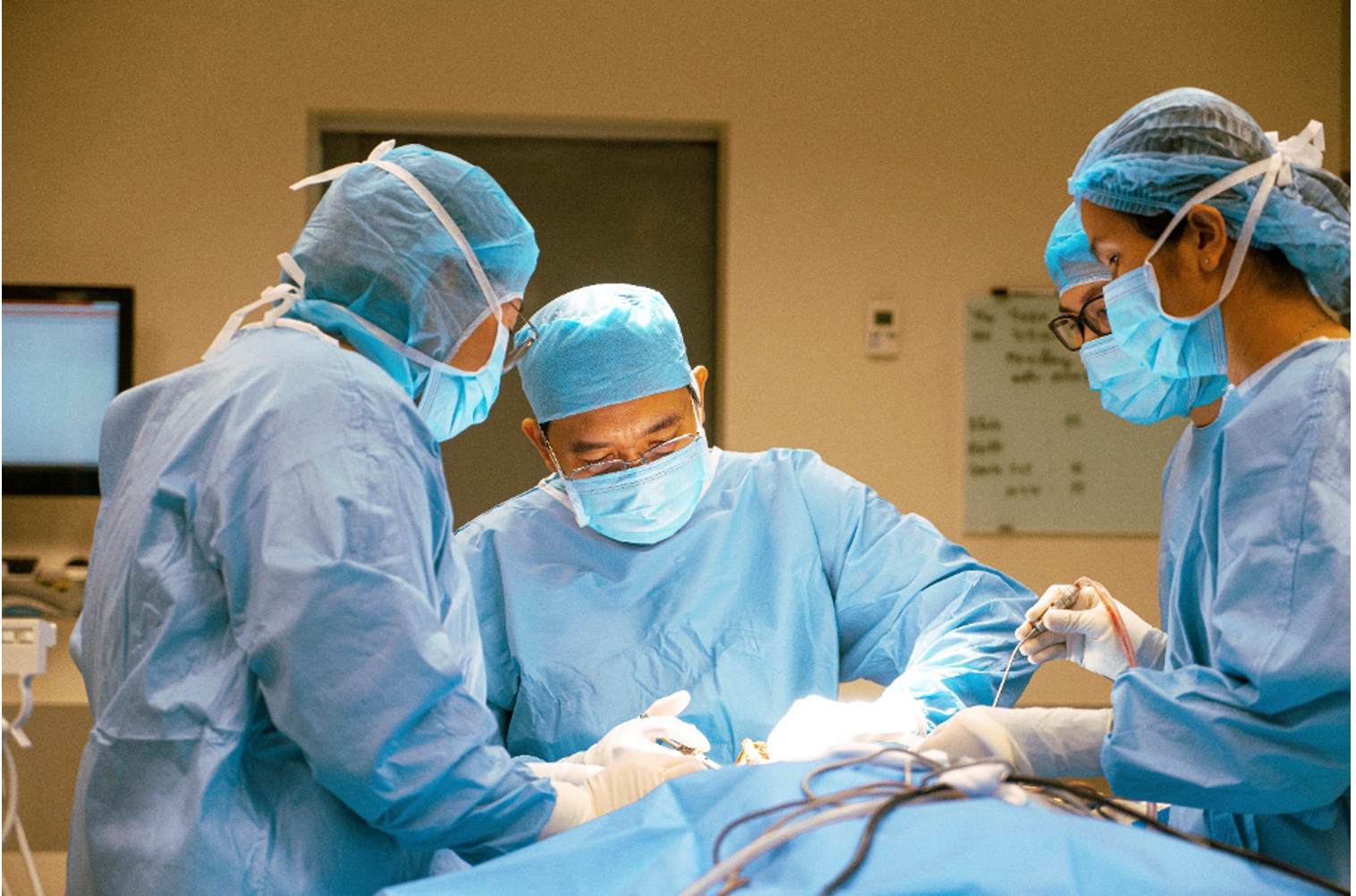
After surgery, Mr. Vasiluk was instructed on how to care for the cyst cavity, perform daily irrigation, and attend regular follow-up appointments to monitor his recovery.
One month later, he returned to FV Hospital. Follow-up imaging showed that the affected area was healing well, with a thin layer of new bone beginning to form. If the bone regeneration process continues favorably, the doctor expects to assess whether Mr. Vasiluk can proceed with further treatment after one to two years.
Dividing treatment into two stages is not uncommon for large jawbone cysts. Decompression is usually performed first, followed by complete removal of the cyst once the lesion has shrunk and the bone has improved. The timing of subsequent treatment depends on the type and size of the lesion, as well as each patient’s response to treatment and bone regeneration.
“Dr. Tung is very skilled, and everything went according to plan,” Mr. Vasiluk said. “Most importantly, he found a way to save my teeth. At every follow-up visit, he told me that the lesion was healing well, and I was very happy.”
Don’t wait until pain appears to discover a jawbone lesion
According to Dr. Tung, Mr. Vasiluk’s case shows that some jawbone lesions can progress for a long time without causing obvious symptoms.
Therefore, patients with impacted or misaligned teeth, or other abnormalities involving the teeth and jaw, should consult a dental and maxillofacial specialist and undergo X-rays when recommended by a doctor. X-ray imaging can help detect lesions deep within the bone that may not be visible during a routine examination.
For large lesions, doctors need to consider their size and location, as well as their impact on the bone, teeth, and surrounding structures, when selecting an appropriate treatment approach. In some cases, staged treatment may help reduce the invasiveness of subsequent surgery and better preserve vital structures.
At FV Hospital, the Maxillofacial and Dental Surgery Department provides diagnosis and treatment for a range of dental and maxillofacial conditions, including complex lesions that require a combination of clinical examination and diagnostic imaging to develop an appropriate treatment plan.
To learn more about specialized dental and maxillofacial treatment services, please contact the Maxillofacial and Dental Surgery Department at FV Hospital, 6 Nguyen Luong Bang, Tan My Ward (formerly District 7), Ho Chi Minh City, or call (028) 3511 3333.







 Google translate